The Fascinating World of Connectomics: Understanding Language Processing
- BrainSightAI Team
- Jun 2, 2023
- 5 min read
Updated: Jun 2, 2023
It feels natural and easy, but in reality, our brains run hundreds of miles per minute to keep up with the flow of conversation. In this blog, we will understand the complexity of language processing through connectomics.
Our brain's receptors record what's being said and convey it to the pathways that carry information to a different cognitive part, which will then process the input. Memory - both of the language and of the topic of discussion - plays a key role here. Then, we formulate a reply. Meanwhile, your brain is also processing emotional responses to the conversation. It could also be thinking, planning, and maybe even having multiple conversations simultaneously!
But this couldn't be possible without the speed and accuracy of those neural connections, conveying information lightning-quick and without pause. Indeed there are so many components in generating a sentence. Connectomics helps us unveil the secrets of language processing. It's a fancy word at first glance, but it's much more straightforward and far more complex than it seems. It helps us explore the neural connections behind our brain's linguistic abilities.
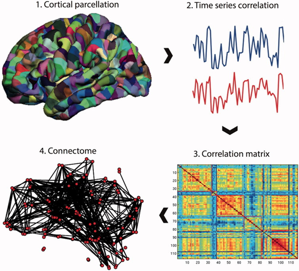
In short, connectomics is the study of structural and functional brain connections. It explains how neurons in the brain and nervous system are connected and interact with each other to produce behaviour and cognition. The total of all these connections is called the "connectome." Structurally, these connections exist in the white matter of the brain.

Connectomics borrows the suffix "-omics" from the word "genomics," which is the study of genomes. Genomics uses "big data" analysis to study DNA sequences, using computational and statistical methods to decode and understand it - the enormous amounts of data giving it the "big data" name. Similarly, connectomics aims to analyze the vast datasets produced by functional and structural brain imaging.
Generally, there are two types of connectomes:
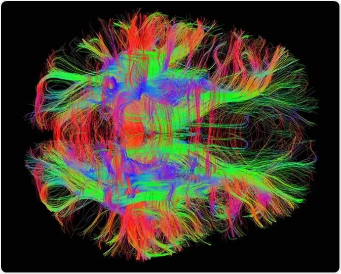
Macroscale connectomes - As the name suggests, the brain is examined on a much larger scale and often as a whole, using the white matter and functions of the brain through MRI techniques. Macroscale connections are pathways in the brain created by bundles of nerve fibers. Thus, macroscale connectomes help us understand how connections work between parts of the brain. dMRI images, like the one below, display white matter to give us an idea of the structural connections. On the other hand, fMRI images are aptly named - they help us learn about functional connections by measuring cerebral blood flow in the brain, which is a marker of neuronal activation.
Microscale connectomes - These connectomes function on a much more microscopic level. They help us study the connections within the subdivided areas of the brain that carry information back and forth. We often take these using electron microscopy.

A brief history of Brain Mapping
In 1909, German anatomist Korbinian Brodmann injured the pride of several cartographers by releasing a map of an area no explorer had set foot in - the brain. Brodmann had spent months observing neurons under a microscope using Nissl staining, studying their cell structure in a method called cytoarchitecture (cyto = cell). Brodmann's map divided the brain into 52 different areas based on their structures, numbered from 1 to 52, which dealt with different functions. For example, Area 17 is the primary visual cortex, and Area 4 is the primary motor cortex.
Brodmann's map, pictured below, was a huge step forward for neuroscientists. The Brodmann areas have since been subdivided into smaller parts, each with more specific functions, as we learn more about the brain.

Many attempts followed to understand brain connectivity and function better. One of the more significant breakthroughs was the first (and only so far) fully reconstructed connectome, that of a roundworm Caenorhabditis elegans, in 1991. At the time, the authors called it a neural circuitry database. This work by Achacoso and Yamamoto was based on the first electron micrographs of Caenorhabditis elegans produced by White, Brenner et al. in 1986.
In 2009, the Human Connectome Project (HCP) decided to continue Brodmann's work. Researchers at the HCP spent the next five years both building on and taking apart his brain map to create an entirely new one, which aimed to map the macroscale connections of the human brain. Their new map, pictured below, focuses on creating distinct brain areas based on their functional roles. It also helps us visualize how these functionally similar areas are structurally connected.

While novel and still highly important, Brodmann's approach was more one-dimensional. So the HCP researchers decided to take a more multi-layered approach. First, they categorized areas of the brain based on different factors like cortical architecture, function, functional connectivity, topography, and anatomy. This led to classifying a whopping 180 areas per brain hemisphere, for a total of 360 areas - more than three times Brodmann's original 52.
Applications of Connectomics
By combining the structural and functional connections of the brain, we can create the most comprehensive brain maps in human history. Some breakthroughs of the HCP model's success are:
New models for brain disorders: The ability to study the brain in this new way, combining different types of scans and focusing on connections, allows us to find biomarkers to better detect and treat diseases like Alzheimer's disease
Using graph theory/computational network models: The nodal structure of the HCP brain map helps us visualize and study the brain using models like neural networks
Brain network organization: We can bring anatomy back into the fold by observing what physical areas correspond to what functional areas and their interconnections through the nodal model
Current Connectomics Research
The HCP's brain map was only the beginning, as a launch point for our current study and perception of connectomics. Its application to medicine is almost limitless; for example, it could help us deepen our biological understanding of mental illness. Mental illness does not manifest with a structural biomarker, so it doesn't appear on a regular MRI scan. Instead, using a more functional approach, we can study if there are any anomalies in the functional connectivity of the brain, searching for a 'functional biomarker.' This new perspective could help us better understand the mechanisms that lead to mental disorders.
Connectomics also has boundless potential beyond medicine. For example, we could use it in psychology, anatomy, and physiology to better understand the connection between our brain and conscious mind. It might even help find ways to enrich and improve cognitive function non-invasively through careful analysis of an individual's brain connections.
In September 2020, Abbott et al. published a paper discussing an ambitious project to map a mouse brain at the level of synapses. The authors talk about the influential work of White, Brenner et al. in creating electron microscopy images of multiple thin slices of the brain of a worm and painstakingly tracing each neuron's branches and synaptic connections with other neurons to produce the structure of the worm's nervous system. They assert that mapping a mouse brain connectome would be a transformative project "to apply new and emerging tools to revolutionize our understanding of brain circuits." This ambitious project has multiple aims:
To create an "unbiased catalog" of cells and their synaptic connections
To better understand connections and projections in the mouse brain, from existing light microscopy data
To learn the structure of long-term memory in mammals
To create a path toward describing the neuropathology of brain disorders
To be a launch point for designing non-biological thinking systems
All in all, connectomics is a rapidly growing field that will remain relevant for many years to come. Who knows where it will lead to? For now, we will follow old and new pathways - both literally and figuratively - to try and find out.
Step into the fascinating world of connectomics and unlock the secrets of the brain's intricate connections with BrainSightAI. Join us on this journey of discovery to better understand how our minds work and pave the way for breakthroughs in medicine, psychology, and beyond. Read our other blog on the subject here!
Citations
Michael Sughrue, M. (2022, December 05). What is Connectomics? Retrieved April 23, 2023, from https://www.o8t.com/blog/connectomics
Connectomics. (2023, January 30). Retrieved April 23, 2023, from https://en.wikipedia.org/wiki/Connectomics
Human Connectome Project (HCP). (n.d.). Retrieved April 23, 2023, from https://www.nimh.nih.gov/research/research-funded-by-nimh/research-initiatives/human-connectome-project-hcp
Chen, P., & Flint, J. (n.d.). What Connectomics can learn from genomics. Retrieved April 23, 2023, from https://journals.plos.org/plosgenetics/article?id=10.1371%2Fjournal.pgen.1009692#pgen.1009692.ref001
Abbott, L. F., & Bock, D. D. (2020, September 17). The Mind of a Mouse. Retrieved April 23, 2023, from https://www.sciencedirect.com/science/article/pii/S0092867420310011


Comments